Contents
- 🔍 Introduction to Immunofluorescence
- 🧬 The History of Immunofluorescence
- 🔬 The Principle of Immunofluorescence
- 📸 Fluorophores and Their Applications
- 👩🔬 Immunofluorescence Techniques and Protocols
- 🔬 Instrumentation and Equipment
- 📊 Data Analysis and Interpretation
- 👨⚕️ Applications in Disease Diagnosis and Research
- 💡 Future Directions and Emerging Trends
- 📚 Conclusion and References
- 🤝 Limitations and Challenges
- 🌟 Perspectives and Controversies
- Frequently Asked Questions
- Related Topics
Overview
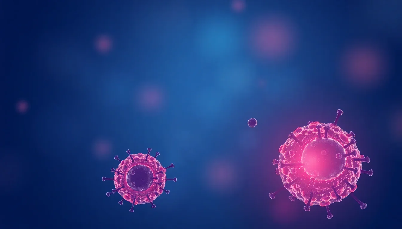
Immunofluorescence is a widely used technique in molecular biology for detecting and localizing biomolecules such as proteins, antibodies, and nucleic acids. Developed in the 1940s by Albert Coons, this method combines the specificity of immunological reactions with the sensitivity of fluorescence microscopy. By using fluorescent dyes attached to antibodies, researchers can visualize the distribution and localization of biomolecules within cells and tissues. With a Vibe score of 8, immunofluorescence has revolutionized the field of cell biology, enabling scientists to study cellular structures and processes in unprecedented detail. However, the technique is not without its limitations, including the potential for non-specific binding and photobleaching. As the field continues to evolve, new advancements in imaging technology and probe development are expected to further enhance the capabilities of immunofluorescence, with potential applications in disease diagnosis and drug development.
🔍 Introduction to Immunofluorescence
Immunofluorescence is a powerful technique used in Biotechnology to visualize and study the distribution of biomolecules within cells and tissues. This method has revolutionized the field of Cell Biology and has numerous applications in Disease Diagnosis and Medical Research. The principle of immunofluorescence is based on the specific binding of Antibodies to their corresponding Antigens. By conjugating these antibodies with fluorescent dyes, researchers can detect and visualize the target molecules with high sensitivity and specificity. For example, Fluorescent Microscopy can be used to study the expression of specific proteins in cells, while Flow Cytometry can be used to analyze the expression of surface markers on cells.
🧬 The History of Immunofluorescence
The history of immunofluorescence dates back to the 1940s, when Albert Coons and his colleagues first developed the technique. Since then, immunofluorescence has undergone significant advancements, with the introduction of new Fluorophores and Imaging Techniques. Today, immunofluorescence is a widely used technique in Molecular Biology and Biochemistry laboratories around the world. The development of Monoclonal Antibodies has also played a crucial role in the advancement of immunofluorescence, allowing for the specific detection of target molecules. For more information on the history of immunofluorescence, see Immunofluorescence History.
🔬 The Principle of Immunofluorescence
The principle of immunofluorescence is based on the specific binding of antibodies to their corresponding antigens. This binding is highly specific, allowing researchers to detect and visualize the target molecules with high sensitivity and specificity. The antibodies used in immunofluorescence are typically conjugated with fluorescent dyes, such as Fluorescein or Rhodamine, which emit light at specific wavelengths. This allows researchers to visualize the target molecules using Fluorescent Microscopy or other imaging techniques. For example, Confocal Microscopy can be used to study the distribution of proteins within cells, while Super Resolution Microscopy can be used to study the distribution of molecules at the nanoscale.
📸 Fluorophores and Their Applications
Fluorophores are the fluorescent dyes used in immunofluorescence to detect and visualize the target molecules. There are many different types of fluorophores available, each with its own unique properties and applications. For example, Green Fluorescent Protein (GFP) is a popular fluorophore used in Cell Biology research, while Red Fluorescent Protein (RFP) is used in Protein-Protein Interactions studies. The choice of fluorophore depends on the specific application and the properties of the target molecule. For more information on fluorophores, see Fluorophores.
👩🔬 Immunofluorescence Techniques and Protocols
Immunofluorescence techniques and protocols vary depending on the specific application and the type of sample being used. For example, Immunofluorescence Staining is commonly used to detect and visualize proteins in cells and tissues, while Flow Cytometry is used to analyze the expression of surface markers on cells. The choice of technique and protocol depends on the specific research question and the properties of the target molecule. For example, Western Blotting can be used to detect and quantify proteins in cells, while ELISA can be used to detect and quantify proteins in serum or other bodily fluids.
🔬 Instrumentation and Equipment
Instrumentation and equipment are critical components of immunofluorescence. Fluorescent Microscopy is the most common imaging technique used in immunofluorescence, and there are many different types of microscopes available, each with its own unique features and applications. For example, Confocal Microscopy is used to study the distribution of proteins within cells, while Super Resolution Microscopy is used to study the distribution of molecules at the nanoscale. The choice of microscope depends on the specific application and the properties of the target molecule. For more information on instrumentation and equipment, see Instrumentation and Equipment.
📊 Data Analysis and Interpretation
Data analysis and interpretation are critical steps in immunofluorescence. The data generated from immunofluorescence experiments can be complex and require specialized software and expertise to analyze and interpret. For example, Image Analysis software can be used to quantify the expression of proteins in cells, while Statistical Analysis software can be used to compare the expression of proteins between different samples. The choice of software and analysis technique depends on the specific research question and the properties of the target molecule. For more information on data analysis and interpretation, see Data Analysis and Interpretation.
👨⚕️ Applications in Disease Diagnosis and Research
Immunofluorescence has numerous applications in disease diagnosis and research. For example, Cancer Research uses immunofluorescence to study the expression of tumor markers and to develop new diagnostic tests. Neurodegenerative Diseases research uses immunofluorescence to study the distribution of proteins in the brain and to develop new therapeutic strategies. The choice of application depends on the specific research question and the properties of the target molecule. For more information on applications, see Applications.
💡 Future Directions and Emerging Trends
Future directions and emerging trends in immunofluorescence include the development of new fluorophores and imaging techniques. For example, Single Molecule Localization Microscopy (SMLM) is a new imaging technique that allows researchers to study the distribution of molecules at the nanoscale. Super Resolution Microscopy is another emerging trend that allows researchers to study the distribution of molecules at the nanoscale. The development of new fluorophores and imaging techniques will continue to advance the field of immunofluorescence and enable new applications and research questions to be addressed.
📚 Conclusion and References
In conclusion, immunofluorescence is a powerful technique used in biotechnology to visualize and study the distribution of biomolecules within cells and tissues. The principle of immunofluorescence is based on the specific binding of antibodies to their corresponding antigens, and the choice of fluorophore and imaging technique depends on the specific application and the properties of the target molecule. For more information on immunofluorescence, see Immunofluorescence.
🤝 Limitations and Challenges
Limitations and challenges of immunofluorescence include the potential for Background Fluorescence and Photobleaching. Background fluorescence can be reduced by using Blocking Agents and optimizing the imaging conditions. Photobleaching can be reduced by using Photostable Fluorophores and optimizing the imaging conditions. The choice of fluorophore and imaging technique depends on the specific application and the properties of the target molecule. For more information on limitations and challenges, see Limitations and Challenges.
🌟 Perspectives and Controversies
Perspectives and controversies in immunofluorescence include the debate over the use of Animal Models in research. Some researchers argue that animal models are necessary for understanding human disease, while others argue that alternative models, such as Cell Culture, should be used instead. The choice of model depends on the specific research question and the properties of the target molecule. For more information on perspectives and controversies, see Perspectives and Controversies.
Key Facts
- Year
- 1941
- Origin
- Harvard University
- Category
- Biotechnology
- Type
- Biological Technique
Frequently Asked Questions
What is immunofluorescence?
Immunofluorescence is a technique used in biotechnology to visualize and study the distribution of biomolecules within cells and tissues. It is based on the specific binding of antibodies to their corresponding antigens, and the choice of fluorophore and imaging technique depends on the specific application and the properties of the target molecule. For more information on immunofluorescence, see Immunofluorescence.
What are the applications of immunofluorescence?
Immunofluorescence has numerous applications in disease diagnosis and research. For example, Cancer Research uses immunofluorescence to study the expression of tumor markers and to develop new diagnostic tests. Neurodegenerative Diseases research uses immunofluorescence to study the distribution of proteins in the brain and to develop new therapeutic strategies. The choice of application depends on the specific research question and the properties of the target molecule. For more information on applications, see Applications.
What are the limitations and challenges of immunofluorescence?
Limitations and challenges of immunofluorescence include the potential for Background Fluorescence and Photobleaching. Background fluorescence can be reduced by using Blocking Agents and optimizing the imaging conditions. Photobleaching can be reduced by using Photostable Fluorophores and optimizing the imaging conditions. The choice of fluorophore and imaging technique depends on the specific application and the properties of the target molecule. For more information on limitations and challenges, see Limitations and Challenges.
What is the future of immunofluorescence?
The future of immunofluorescence includes the development of new fluorophores and imaging techniques. For example, Single Molecule Localization Microscopy (SMLM) is a new imaging technique that allows researchers to study the distribution of molecules at the nanoscale. Super Resolution Microscopy is another emerging trend that allows researchers to study the distribution of molecules at the nanoscale. The development of new fluorophores and imaging techniques will continue to advance the field of immunofluorescence and enable new applications and research questions to be addressed.
What are the perspectives and controversies in immunofluorescence?
Perspectives and controversies in immunofluorescence include the debate over the use of Animal Models in research. Some researchers argue that animal models are necessary for understanding human disease, while others argue that alternative models, such as Cell Culture, should be used instead. The choice of model depends on the specific research question and the properties of the target molecule. For more information on perspectives and controversies, see Perspectives and Controversies.
What is the role of immunofluorescence in disease diagnosis?
Immunofluorescence plays a critical role in disease diagnosis by allowing researchers to visualize and study the distribution of biomolecules within cells and tissues. For example, Cancer Diagnosis uses immunofluorescence to study the expression of tumor markers and to develop new diagnostic tests. Neurodegenerative Diseases Diagnosis uses immunofluorescence to study the distribution of proteins in the brain and to develop new diagnostic tests. The choice of application depends on the specific research question and the properties of the target molecule. For more information on disease diagnosis, see Disease Diagnosis.
What is the role of immunofluorescence in medical research?
Immunofluorescence plays a critical role in medical research by allowing researchers to visualize and study the distribution of biomolecules within cells and tissues. For example, Cancer Research uses immunofluorescence to study the expression of tumor markers and to develop new therapeutic strategies. Neurodegenerative Diseases Research uses immunofluorescence to study the distribution of proteins in the brain and to develop new therapeutic strategies. The choice of application depends on the specific research question and the properties of the target molecule. For more information on medical research, see Medical Research.